Key Points
- The spinal cord extends from the brainstem in the foramen magnum to the conus medullaris at L1 in adults or L3 in newborns.
- The anterior spinal artery supplies the anterior two-thirds of the spinal cord and medulla, while the posterior spinal arteries supply the remaining one-third of the spinal cord.
- The anterior spinal cord is more prone to ischemia and can lead to anterior spinal artery syndrome.
Anatomy
- The spinal cord is an extension of the brainstem spanning from the foramen magnum to the conus medullaris as the filum terminus at L1 in adults, after which it is referred to as the cauda equina (L2-sacral).1-3 In the newborn, the conus medullaris extends to L3.
- The spinal cord is a tubular structure that, on cross-section, has an elliptical shape in the cervical region, and a round shape in the thoracic region.2
- The spinal cord is thickest in two areas: a cervical enlargement (C5-T1) that innervates the upper extremities, and a smaller lumbar enlargement (T9-L2) that innervates the lower extremities.2
- The spinal cord can be divided into 31 segments: 8 cervical, 12 thoracic, 5 lumbar, 5 sacral, and 1 coccygeal segment. Except for C1, which has no sensory nerve root, each segment has a pair of dorsal (sensory) and ventral (motor) nerve roots that join to form a mixed spinal nerve that exits the vertebral canal through the intervertebral foramina.1-3
- Spinal nerves in the cervical region exit above their corresponding vertebrae. Starting with T1, the spinal nerves exit below their corresponding vertebrae.1
- The lowermost nerve roots (L2-5, S1-5, and S1-5) fan out distally from the lower part of the spinal cord to form the cauda equina (Latin for horse’s tail).3

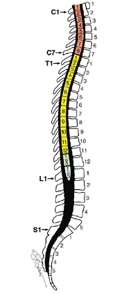
Figure 1. Relationship between the spinal cord and vertebral bodies. Image used with permission from Loo NH, Matchett G. Use of a tilting orthopedic fracture table to facilitate proper patient positioning during intrathecal neurolysis with hyperbaric phenol. AA Case Rep. 2017; 9(16):164-68.4
Cross-Sectional Anatomy
- The spinal cord is composed of longitudinal columns of nuclei (gray matter) surrounded by ascending and descending tracts (white matter).1-3
- The gray matter is butterfly or H-shaped and is divided into anterior (ventral), posterior (dorsal), and lateral (intermediate) horns.
- The anterior horns contain alpha and gamma motor neurons and interneurons.
- Somatic motor neurons travel in the medial motor columns, where they innervate the axial muscles of the body, or in the lateral motor columns, where they innervate the upper and lower limbs.
- The posterior horns are where somatic and visceral afferent (sensory) fibers enter the spinal cord.
- The lateral horns contain the autonomic preganglionic cells. The preganglionic sympathetic neurons are located from C8-L1, and the preganglionic parasympathetic neurons are located from S2-S4.
- The gray matter horns divide the white matter into posterior (dorsal), lateral, and anterior (ventral) columns (Figure 2).1-3
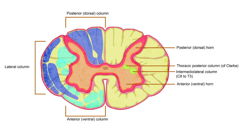
Figure 2. Cross-sectional anatomy of the spinal cord. The posterior, lateral, and anterior columns of white matter are illustrated on the left side. Ascending pathways are colored dark blue, and descending pathways are colored light blue. The posterior, intermediate, and ventral horns of the gray matter are illustrated on the right side. Redrawn from Cho TA. Spinal cord functional anatomy. Continuum (Minnea Minn). 2015;21(1): 13-35.1
- The white matter columns include both ascending and descending tracts.1-3 Ascending neural tracts convey information from the periphery to the brain, while descending tracts convey information from the brain to the periphery (Figure 3).
- Major ascending tracts include:
- dorsal column: conveys tactile sensation, vibration, and proprioception;
- spinothalamic tract: conveys pain, temperature, and crude touch.
- Major descending tracts include:
- corticospinal tract: controls fine motor movement.
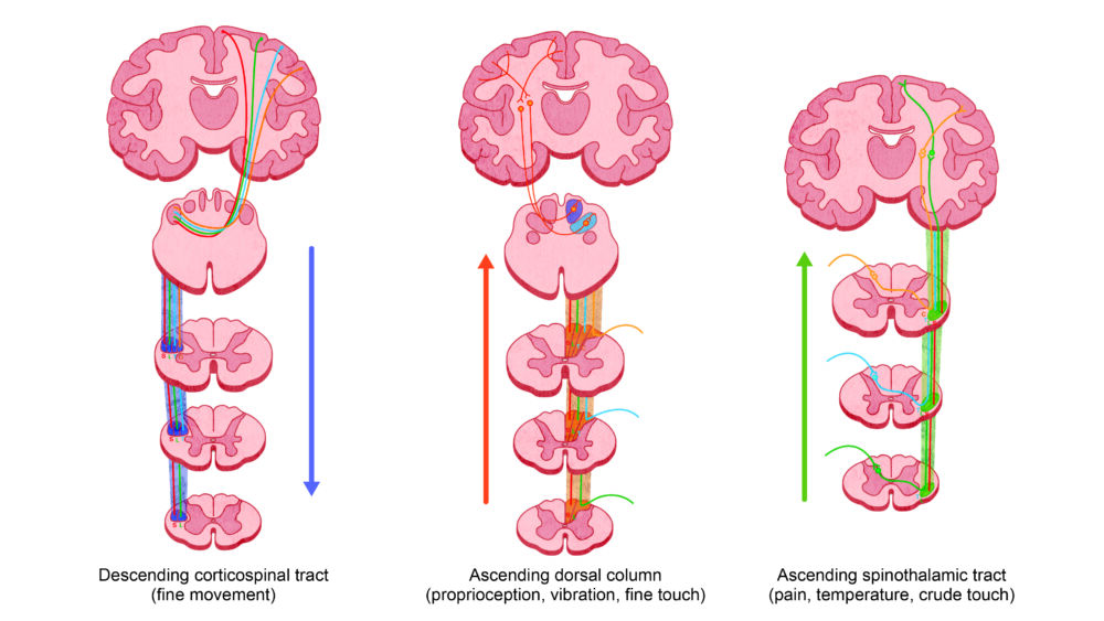
Figure 3. Major ascending and descending tracts of the spinal cord. Redrawn from Kunam VK, et al. Incomplete cord syndromes. Clinical and imaging review. Radiographics. 2018; 38: 1201-22.2
Blood Supply
- The anterior spinal artery originates from the vertebral artery and supplies the anterior two-thirds of the spinal cord and medulla (Figure 4).1-3
- The upper cervical segment of the spinal cord receives most of its blood supply from the vertebral arteries. The thoracic portion of the anterior spinal artery is supplied by the anterior radicular arteries: 1 or 2 cervical, 2-3 thoracic, and 1-2 lumbar.5
- The great radicular artery, also known as the Artery of Adamkiewicz, is the largest radicular artery and is the major blood supply to the lower two-thirds of the spinal cord. It is generally located on the left side between T9-T12 (75% of cases), though it can be located anywhere between T7-L4 (Figure 4).5
- Two posterior spinal arteries (originating from the inferior cerebellar artery), as well as the segmental spinal arteries (originating from the intercostal and lumbar arteries) supply the posterior one-third of the spinal cord.1-3
- Similar to the arterial supply, there are three longitudinal anterior spinal veins and three posterior spinal veins that drain into the internal vertebral venous plexus in the medial and lateral components of the epidural space.
- The anterior and deep portions of the spinal cord are more prone to ischemia due to fewer anterior medullary feeder vessels than the posterior region and can lead to anterior spinal artery syndrome, characterized by ischemia of the bilateral corticospinal and spinothalamic tracts. This causes loss of motor function and loss of pain and temperature sensation below the level of the lesion. Common etiologies include aortic dissection, emboli, covering of the large segments of the thoracoabdominal aorta with endovascular stents and surgical dissection during thoracoabdominal aortic aneurysm repair.
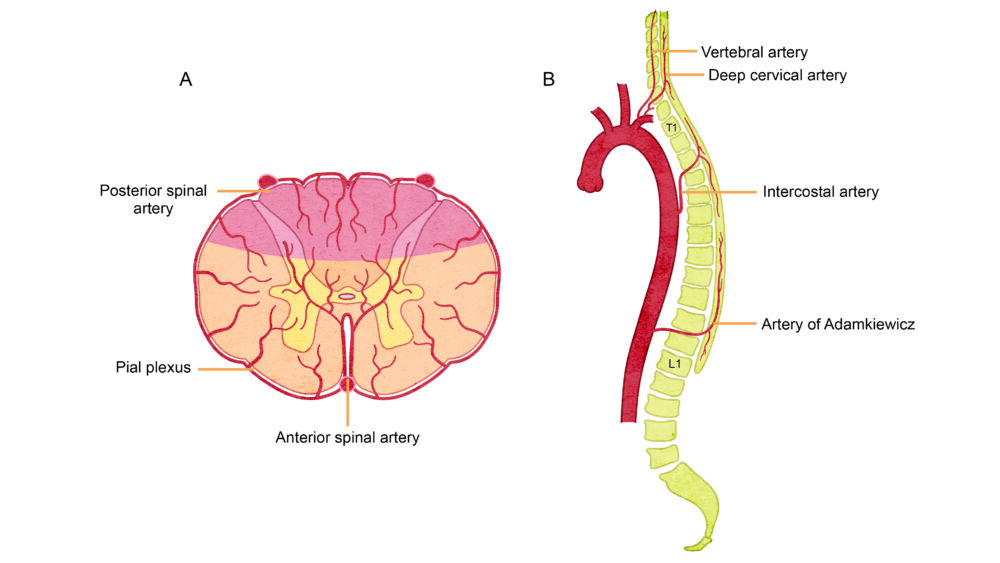
Figure 4. Blood supply of the spinal cord. A- A single anterior spinal artery supplies the anterior two-thirds of the spinal cord, and paired posterior spinal arteries supply the posterior one-third of the spinal cord. B- The artery of Adamkiewicz is the largest radicular artery that usually arises from the left side of the aorta between T9 and L2 and provides the primary blood supply to the lumbar and sacral segments. Redrawn from. Kunam VK, et al. Incomplete cord syndromes. Clinical and imaging review. Radiographics. 2018; 38: 1201-22.2
References
- Cho TA. Spinal cord functional anatomy. Continuum (Minnea Minn). 2015;21(1): 13-35. PubMed
- Kunam VK, Velayudhan V, Chaudhry ZA, et al. Incomplete cord syndromes. Clinical and imaging review. Radiographics. 2018; 38: 1201-22. PubMed
- Hardy TA. Spinal cord anatomy and localization. Continuum (Minnea Minn). 2021; 27(1):12-29. PubMed
- Loo NH, Matchett G. Use of a tilting orthopedic fracture table to facilitate proper patient positioning during intrathecal neurolysis with hyperbaric phenol. AA Case Rep. 2017; 9(16): 164-68. PubMed
- Shalabi A, Chang J. Anesthesia for vascular surgery. In: Miller’s Anesthesia. 6th edition. Elsevier. 2020: 1825-67.
Copyright Information

This work is licensed under a Creative Commons Attribution-NonCommercial-NoDerivatives 4.0 International License.